The Anthropology Tutorials website will be taken offline on January 9, 2026.
Due to federal accessibility requirements taking effect in 2026, we will be removing this site. If you use materials from this site in your courses or research, please seek alternative resources.
Molecular
Level of Genetics
So far in
this tutorial, chromosomes and genes have been described broadly without saying precisely
what they are composed of and how they function. In order to better understand them,
we need to look at the molecular level of the cell.
Water is by far the most common type of
molecule in the human body, normally amounting to 50-60% of our total adult
weight. Most of the
remaining molecules fall into one of four categories:
|
1. |
carbohydrates
 (sugars and starches) (sugars and starches) |
|
2. |
lipids
 (fats, oils,
waxes, fatty acids, triglycerides and cholesterol)
 |
|
3. |
proteins
 |
|
4. |
nucleic acids
 |
Proteins are large chain-like molecules that are twisted and folded
back on themselves in complex patterns. They serve as structural material for the
body, gas transporters in blood, hormones
 ,
antibodies
 ,
neurotransmitters
 ,
and enzymes
 .
When looking at someone, you mostly see proteins since skin,
hair, and muscles are primarily made of them. Proteins acting as
enzymes are
particularly important
substances because they trigger and control the chemical reactions by which carbohydrates,
lipids, and other substances are created. Our human bodies produce about 90,000 different kinds of proteins, all of which consist of simpler units called
amino acids
 . Proteins in all organisms are
mostly composed of just 20 kinds of amino acids. Proteins differ in the number,
sequence, and kinds of amino acids. Our bodies create some of these amino acids,
while others come directly from food that we consume.
|
AMINO
ACIDS |
| alanine |
glutamic acid |
leucine |
serine |
| arginine |
glutamine |
lysine |
threonine |
| asparagine |
glycine |
methionine |
tryptophan |
| aspartic acid |
histidine |
phenylalanine |
tyrosine |
| cysteine |
isoleucine |
proline |
valine |
|
(NOTE: The 8 amino acids
in red cannot be
synthesized by the
human body and must be obtained from food.
They are referred
to as "essential amino acids." Arginine and histidine
(shown in
green)
evidently are essential only for infants and are, therefore,
considered to be semi-essential.) |
Proteins, and
subsequently amino acids, are mostly made up of just four elements: carbon, oxygen,
hydrogen, and nitrogen. In fact, 96.3% of a human body is composed of these common
elements.
The largest molecules in
people and other organisms are nucleic acids. Like proteins, they consist of
very long chains of simpler units. However, the components, shapes, and functions of
nucleic acids differ significantly from those of proteins. There are two basic
forms of nucleic acids: DNA (deoxyribonucleic acid
 ) and
RNA (ribonucleic
acid
 ). Both play critical roles in the production of
proteins. Some kinds of RNA perform other critical functions in our
cells not related to protein synthesis.
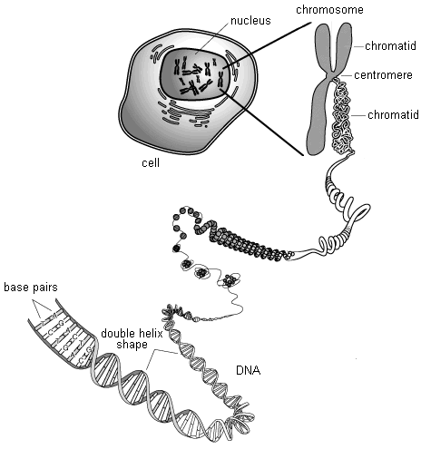
A chromosome
consists mainly of a single very long DNA molecule and proteins called
histones
 that densely package the DNA.
There are many small RNA molecules surrounding the DNA as well. Each of
our DNA
molecules contains the genetic codes, or genes, for the synthesis of many
different kinds of proteins and for the regulation of other genes. In a sense, a
DNA molecule contains a sequence of permanently stored blueprints or recipes
that are used mostly by our cells to assemble proteins out of amino acids.
In other words, chromosomes are packets of information encoded by DNA.
If all of the DNA molecules in a single human cell were straightened out and arranged end to end, it would
form a thread about
6 feet long but only a few atoms across. If the DNA in all of a normal
adult human's cells were arranged in this way, the thread could reach the moon
about 6,000 times.
DNA molecules have the shape of a
double helix
 , which is
like a twisted ladder. The sides of the ladder are composed of sugar and phosphate
units, while the rungs consist of complementary pairs of four different chemical
bases. The base guanine only bonds to cytosine and adenine only bonds to
thymine. Each combined sugar, phosphate, and base subunit is a
nucleotide
 .
.
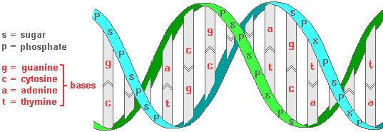 |
Section of a DNA molecule
showing the double helix molecular shape |
The linear
sequence of base pairs along the length of a DNA molecule is the genetic code for the assembly of
particular amino acids to
make specific types of proteins. Therefore, a gene is essentially a
recipe consisting of a sequence of some of these base pairs. The
sequence is usually not
continuous but
is in several different sections of a DNA molecule. Sections of the same
gene can be assembled in different ways resulting in recipes for different
kinds of proteins. This "alternative splicing" process is common,
occurring in 92-94% of human genes.
Only
1.1-1.5% of the approximately 3 billion base pairs in
human DNA actually code for proteins. These meaningful code sequences are called
exons
 . The remaining
98+% of our DNA base pairs were in the past
thought to consist merely of genetic junk, including meaningless code section
duplications and ancient remnants
of parasitic DNA that invaded our ancestors' cells many millions of years ago. However, it is now becoming clear that
much of
this "junk" actually has important functions. These non-protein coding
sections are referred to
as introns
 . At least 80%
of the intron regions of human DNA contain switches to turn the genes on and off.
There are at least 4 million of these switches. Many of them are involved in
monitoring and controlling cell functions throughout the body. Some
also may be involved in a wide range of diseases including autoimmune
responses (multiple sclerosis, lupis, rheumatoid arthritis, Crohn's disease,
and celiac disease).
Textbooks
written before 2001 most often stated that there are 100,000 human genes.
This estimate was significantly reduced to about 30,000 as a result of completion of the
initial phase of the
Human Genome Project in that
year. The number was further revised downward in 2004 by the National
Human Genome Research Institute (NHGRI). It is now known that there are
about 20,500 still
functioning human protein-coding genes.
Since the human genome "parts list"
was compiled, research has largely focused on what these parts do--i.e., what
proteins they code for and what those proteins do in our bodies. In
addition, there is intense ongoing research to understand the short RNA
molecules that are coded for by introns.
 Extract Your Own DNA--simple
how-to instructions to separate out your own DNA at home.
This link takes
you to an external website. To return here, you must click the
"back" button
on your browser program.
(length = 2 mins, 45 secs) |
There is far more DNA variation
between people than was thought possible in 2001 when the human genome
was first worked out. The variations mostly show up in 1500 DNA
regions (about 12% of the total). The differences are commonly in the form of
deletions, duplications, and reversals of DNA segments and individual bases.
Research is still at the beginning of the process of determining the
significance of these differences. Understanding them very likely
will shed considerable light on whether or not individuals might develop
particular diseases. These variations are far more common and
extensive than previously assumed. Current estimates are that
65-80% of people have copy number variants (CNV's) that are at least
100,000 base units long.
About half of the sequences of base units in human
nuclear DNA are
"mobile elements." They move around inserting new copies of themselves.
When these insertions go into a gene, the result is a changed protein recipe.
This is a major source of new genetic variation and potentially a cause of
genetic diseases such as hemophilia and
muscular dystrophy in families
that did not previously have these defective genes.
|
|
|
|
| |
 |
|
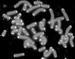
Human chromosomes
with
telomeres shown
in white |
NOTE:
At both ends of chromosomes there are telomere caps, which are
sections of several thousand repeat DNA base unit sequences that are not
part of genes (TTAGGG, TTAGGG, TTAGGG, etc.).
Telomeres help protect critical DNA sequences from being changed by
recombination. However, the telomeres become slightly shorter every
time cell division occurs. When they have only around 77 base units
remaining, they become unstable, leading to the fusion of chromosomes and
cell death. This is a normal consequence of aging. Very short telomeres are also associated with early stages
of cancer.
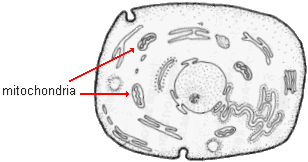
Not all of our DNA is in the
cell nucleus. A small amount is in a circular looping chromosome in
mitochondria
 --organelles located in the cytoplasm. Mitochondria use glucose, a
sugar, derived from the
food we eat to produce adenosine triphosphate (ATP). This is the fuel for many cell functions.
Without ATP, cell activity would cease and we would die. There are about 500 mitochondria in each
human
cell. Muscle cells have more because they need additional ATP to
function. The 16,569 base units of human mitochondrial DNA (mtDNA) have only 37 genes
of which 13 code for proteins.
 |
|
Generalized
animal cell |
|
|
|
|
|
 |
| |
Sperm cell |
Unlike nuclear DNA (nDNA) in
chromosomes, mtDNA is
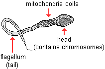
inherited almost exclusively from our mothers. In sperm
cells, the mitochondria are located in coils between the flagellum tail and
the head. They provide the fuel that allows sperm to move. It has
been widely assumed that, at
conception, the flagellum along with the mitochondria do not
enter the ovum. We now know that this is not always true in the case
of humans and other mammals. However, less than .1%
of the mitochondrial DNA in a zygote normally comes from the father, and the
genes in the paternal mtDNA apparently get turned off. Subsequently, for all intent purposes, we only inherit maternal mtDNA.
The second
type of nucleic acid, RNA, consists of molecules that are single stranded copies of
nuclear DNA segments. They are smaller than DNA molecules and do not have the double
helix shape. In addition, the DNA base thymine is replaced by the base uracil
in RNA molecules. The sugar component is also somewhat different. RNA is found in both
the cell nucleus and the cytoplasm. The different kinds of RNA are
described below.
Protein Synthesis
The genetic code is central to two critical functions
within our cells. They are the creation of new proteins and the
duplication of chromosomes during cell division. The former process is
actually an assembly of existing amino acids in their proper sequences for
each type of protein. Subsequently, it is referred to as protein
synthesis rather than production. In this process, the recipe for a
needed protein is first copied. Specifically, the relevant DNA code
sequence is transcribed, or copied, to RNA. The
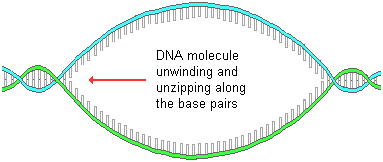
process begins by a section of a DNA molecule unwinding and then unzipping in response to
several interacting enzymes. The separation occurs between the bases, as shown below.
|
DNA molecule
partially
unwinding and unzipping |
|
 |
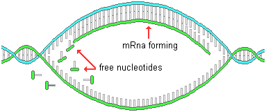
Free nucleotides in the nucleus are attracted to
complimentary bases on the exposed DNA strands (guanine to
cytosine and adenine to thymine).
The result
of this sequential bonding of base units is the formation of a
messenger RNA (mRNA) molecule that is a copy, or
transcription, of specific
sections of the nuclear DNA molecule corresponding to a gene. Many identical copies
are made, one right after another. In the process of mRNA formation, the
non-coding sections are removed and the remaining exons are spliced
together.
|
Free nucleotides attracted
to
exposed bases of a
partially unzipped DNA
molecule |
|
 |
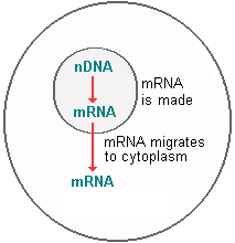
These new identical
messenger RNA molecules then leave the nucleus and go out into the cytoplasm where the
protein they are coded for is actually synthesized or assembled.
|
mRNA migrating out
of the cell
nucleus |
|
 |
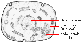
Specifically,
the messenger RNA molecules migrate from the chromosomes to the
ribosomes
 , which are small granule-like
organelles
in the cytoplasm. Some ribosomes are on the surface of long membrane
networks called endoplasmic reticula
 ,
while others are free ribosomes.
Assembly of
proteins takes place at the site of the ribosomes.
|
Generalized animal cell |
|
 |
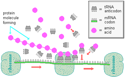
Protein synthesis begins as
ribosomes move along the messenger RNA strand and attach
transfer RNA (tRNA) anticodons
 (each with 3
bases) to triplets of complementary bases on the mRNA.
 |
Protein
synthesis at the ribosomes initiated by mRNA momentarily bonding with tRNA
(This schematic representation is a
simplification of the actual shapes and processes.) |
Each transfer RNA attracts and brings a
specific amino acid along with it. As a ribosome translates (or
activates) the
messenger RNA code, a protein chain is assembled, one amino acid at a time.
Each kind of amino acid has a particular codon that specifies it. A codon is a
sequence of three nucleotide components chemically bound together (illustrated below).
As mentioned earlier, every nucleotide consists of a sugar, a phosphate, and a base. Codons differ in terms of the sequence of their three bases. For example, the sequence CAG (cytosine-adenine-guanine) is a code for the amino acid glutamine.
 |
Sugar-phosphate-base chemical
bond of a codon
(Simplified representation
rather than actual molecular shape) |
This genetic code permits 64 different codons because each of the 3
nucleotides can have 1 of the 4 bases ( 4 X 4 X 4 = 64 ). Because there are
many fewer
than 64 amino acids, the code system has built in redundancy--most amino
acids can be attracted by transfer RNA having several different base triplet
combinations. In
other words, some codons are functionally equivalent, as shown in the table below. For
instance, asparagine is specified with the mRNA codon sequence AAU
(adenine-adenine-uracil). However, AAC (adenine-adenine-cytosine) also works.
|
DNA
AND RNA
CODES FOR AMINO ACIDS |
| Amino acids |
Corresponding DNA
codons |
Corresponding RNA codons |
| alanine |
CGA, CGG, CGT,
CGC |
GCU, GCC, GCA,
GCG |
| arginine |
GCA, GCG, GCT,
GCC, TCT, TCC |
CGU, CGC, CGA,
CGG, AGA, AGG |
| asparagine |
TTA, TTG |
AAU, AAC |
| aspartic acid |
CTA, CTG |
GAU, GAC |
| cysteine |
ACA, ACG |
UGU, UGC |
| glutamic acid |
CTT,CTC |
GAA, GAG |
| glutamine |
GTT, GTC |
CAA, CAG |
| glycine |
CCA, CCG, CCT,
CCC |
GGU, GGC, GGA, GGG |
| histidine |
GTA, GTG |
CAU, CAC |
| isoleucine |
TAA, TAG, TAT |
AUU, AUC, AUA |
| leucine |
AAT, AAC, GAA,
GAG, GAT, GAC |
UUA, UUG, CUU,
CUC, CUA, CUG |
| lysine |
TTT, TTC |
AAA, AAG |
| methionine
(start codon) |
TAC |
AUG |
| phenylalanine |
AAA, AAG |
UUU, UUC |
| proline |
GGA, GGG, GGT,
GGC |
CCU, CCC, CCA, CCG |
| serine |
AGA, AGG, AGT,
AGC, TCA, TCG |
UCU, UCC, UCA,
UCG, AGU, AGC |
| threonine |
TGA, TGG, TGT,
TGC |
ACU, ACC, ACA, ACG |
| tryptophan |
ACC |
UGG |
| tyrosine |
ATA, ATG |
UAU, UAC |
| valine |
CAA, CAG, CAT,
CAC |
GUU, GUC, GUA, GUG |
| (stop codon) |
ATT, ATC, ACT |
UAA, UAG, UGA |
| NOTE: the DNA base thymine
(T) is replaced with uracil (U) in RNA. |
Not all
codons specify amino acid components to be included in a protein. For instance, a start
codon appears in DNA at the beginning of the code sequence for each gene and a stop
codon is at the end. In other words, they indicate where a protein recipe begins
and ends.
Most plant and animal cells, including those of
humans,
have thousands to millions of ribosomes. Many ribosomes simultaneously translate
identical strands of messenger RNA. As a result, the synthesis of proteins can be
rapid and massive. These same processes usually occur at the same time in millions of
cells when a particular protein is needed. Inevitably, there are some
errors in the code copying process. The result is mutations that cause the creation of
incorrect proteins. However, there is an error checking and correction
process carried out by enzymes that catches many of these errors. Recent
research suggests that DNA to RNA copy mistakes are common.
Most of our genes code for several
different proteins. This should not be surprising since we have only
about a third as many genes as we do kinds of proteins. Some genes act in consort with other
genes to produce the same protein. Some genes do
not
code for proteins at all. Recent research has shown that at least 200-255 of our genes
code for micro RNA (miRNA) molecules instead. These are not messenger RNA's that
transport the recipe for protein synthesis. Rather, they are small RNA
molecules consisting of only around 20-25 base units. They perform
important functions similar to enzymes in regulating chemical reactions in our
cells, especially in the embryonic stage at the beginning of life. It is
thought that at least 1/3 of human genes are controlled in some way by micro-RNA
molecules.
DNA Replication
In addition to keeping the
blueprints for protein synthesis, DNA has one further function. It replicates, or
duplicates, itself. This occurs whenever a chromosome is duplicated for
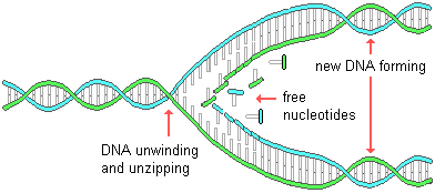
cell division. At the beginning of this process, the DNA molecule of a
stretched out chromosome
unwinds and unzips along its bases beginning at one end. Then in response to an enzyme, free nucleotides
pair up with corresponding bases on both of the DNA strands, as illustrated below.
This results in the formation of two exact copies of the original molecule. Nuclear
DNA replication occurs in the rest period (interphase) just before mitosis and meiosis.
|
DNA
replication
process |
|
 |
Occasionally, an error is
made in DNA replication. For example, an incorrect base pair may be included. This
constitutes a mutation. If it occurs in the
formation of sex cells, the mutation may
be inherited and passed on in future generations. Such errors are the
principal
source of new genetic variation for a species and, subsequently, play a major
role in evolution. DNA replication errors are also responsible for changes in somatic cells
that result in the uncontrolled tumor growths of cancers.
Universality of the
Genetic Code
The DNA code system of humans is not unique but is shared by all living things
on our planet. The same codons code for the same amino acids in people, dogs,
fleas, and even bacteria. In addition, we share many genes with other
creatures. For instance, about 90% of human genes are identical to those
of a mouse. Even more surprising is the fact that more than 1/3 of our
genes are shared with a primitive group of worm species known as nematodes. The universal nature of the genetic code is compelling evidence for the
evolution of all organisms from the same early life forms.
 The Common Genetic Code--evidence
that humans share a common
genetic code with
other organisms.
This link takes
you to an external website. To return here, you must
click the "back" button on
your browser program. (length =
4 mins, 20 secs) |
Epigenome
Whether a gene is copied for protein synthesis and
what the product of that copy becomes is largely determined by signals from
proteins acting as markers and switches along the DNA double helix
structure. This chemical signaling system, referred to as the
epigenome, is very likely as important as the DNA itself in determining
the phenotype of individuals. It is becoming increasingly clear that
epigenetic signals can be altered by the environment. This partly
explains why monozygotic twins progressively become different from one
another as they grow older despite the fact that they are genetically
identical.
Epigenetic changes can be passed on to future
generations because epigenome proteins are inherited along with DNA in
chromosomes from parents. Unlike mutations in DNA sequences, however,
epigenetic alterations potentially can be reversible. For example, if
a change in an epigenetic protein causes a disease, it could be possible to
alter that protein and bring about a cure. As a consequence, learning
more about the human epigenome is likely to be an important area for future
medical research.
 Epigenetics--excerpt from the PBS series Nova Science Now
(July 24, 2007)
This link takes
you to an external website. To return here, you must click the
"back" button
on your browser program.
(length = 13 mins, 2 secs) |
The effects of changes in an individual's epigenome
can be far reaching. Not only can they affect physical appearance and
health, but they can also alter behavior. For instance, the babies of
rat mothers who are unusually nurturing are likely to have altered
expressions of the genes involved with stress responses. These changes allow
them to handle stress better as adults. It is thought that a similar
connection occurs in humans. Likewise, some researchers have suggested
that children of parents who were severely undernourished at the time of
conception have a greater chance of developing Schizophrenia. The
assumption is that famine conditions resulted in epigenome alterations
responsible for this severe mental disorder in future children.
When sperm cells enter an egg at conception,
they bring along with them substantial amounts of paternal RNA and
epigenetic proteins which apparently play an important role in the
development of embryos.
NOTE:
If you had difficulty in understanding these
complex processes, you may wish to go over them again.
Keep in mind that the graphic shapes used here to model the components of
DNA, RNA, protein, and amino acid are not accurate representations of the
real shapes and the relative sizes. You can also review this
information with the animated tutorials on DNA, genes, chromosomes, and
proteins at the Learn Genetics web site linked below:
 Learn
Genetics: Tour of the Basics--animated tutorial showing the
relationship between
DNA, genes,
chromosomes, and proteins. Produced by the Genetics Science
Learning
Center of the University of Utah. This link takes
you to an external website. To return
here, you must click the "back"
button on your browser program. |
NOTE:
There is an RNA-based defense system in the cytoplasm of cells that
functions to prevent the genetic material of invading viruses from being
replicated. This is known as RNA interference
or RNAi. The video linked below provides a simple explanation of how
this system works.
|
 RNAi
explained--excerpt from the PBS series Nova Science Now (July
25, 2005)
This link takes
you to an external website. To return here, you must click the
"back" button
on your browser program.
(length = 15 mins) |
Copyright 1998-2014 by Dennis
O'Neil.
All rights reserved.
illustration credits
![]() ,
antibodies
,
antibodies
![]() ,
neurotransmitters
,
neurotransmitters
![]() ,
and enzymes
,
and enzymes
![]() .
When looking at someone, you mostly see proteins since skin,
hair, and muscles are primarily made of them. Proteins acting as
enzymes are
particularly important
substances because they trigger and control the chemical reactions by which carbohydrates,
lipids, and other substances are created. Our human bodies produce about 90,000 different kinds of proteins, all of which consist of simpler units called
amino acids
.
When looking at someone, you mostly see proteins since skin,
hair, and muscles are primarily made of them. Proteins acting as
enzymes are
particularly important
substances because they trigger and control the chemical reactions by which carbohydrates,
lipids, and other substances are created. Our human bodies produce about 90,000 different kinds of proteins, all of which consist of simpler units called
amino acids
![]() . Proteins in all organisms are
mostly composed of just 20 kinds of amino acids. Proteins differ in the number,
sequence, and kinds of amino acids. Our bodies create some of these amino acids,
while others come directly from food that we consume.
. Proteins in all organisms are
mostly composed of just 20 kinds of amino acids. Proteins differ in the number,
sequence, and kinds of amino acids. Our bodies create some of these amino acids,
while others come directly from food that we consume.![]() ) and
RNA (ribonucleic
acid
) and
RNA (ribonucleic
acid
![]() ). Both play critical roles in the production of
proteins. Some kinds of RNA perform other critical functions in our
cells not related to protein synthesis.
). Both play critical roles in the production of
proteins. Some kinds of RNA perform other critical functions in our
cells not related to protein synthesis.![]() that densely package the DNA.
There are many small RNA molecules surrounding the DNA as well. Each of
our DNA
molecules contains the genetic codes, or genes, for the synthesis of many
different kinds of proteins and for the regulation of other genes. In a sense, a
DNA molecule contains a sequence of permanently stored blueprints or recipes
that are used mostly by our cells to assemble proteins out of amino acids.
In other words, chromosomes are packets of information encoded by DNA.
If all of the DNA molecules in a single human cell were straightened out and arranged end to end, it would
form a thread about
6 feet long but only a few atoms across. If the DNA in all of a normal
adult human's cells were arranged in this way, the thread could reach the moon
about 6,000 times.
that densely package the DNA.
There are many small RNA molecules surrounding the DNA as well. Each of
our DNA
molecules contains the genetic codes, or genes, for the synthesis of many
different kinds of proteins and for the regulation of other genes. In a sense, a
DNA molecule contains a sequence of permanently stored blueprints or recipes
that are used mostly by our cells to assemble proteins out of amino acids.
In other words, chromosomes are packets of information encoded by DNA.
If all of the DNA molecules in a single human cell were straightened out and arranged end to end, it would
form a thread about
6 feet long but only a few atoms across. If the DNA in all of a normal
adult human's cells were arranged in this way, the thread could reach the moon
about 6,000 times.![]() , which is
like a twisted ladder. The sides of the ladder are composed of sugar and phosphate
units, while the rungs consist of complementary pairs of four different chemical
bases. The base guanine only bonds to cytosine and adenine only bonds to
thymine. Each combined sugar, phosphate, and base subunit is a
nucleotide
, which is
like a twisted ladder. The sides of the ladder are composed of sugar and phosphate
units, while the rungs consist of complementary pairs of four different chemical
bases. The base guanine only bonds to cytosine and adenine only bonds to
thymine. Each combined sugar, phosphate, and base subunit is a
nucleotide
![]() .
.
